Figure 4.
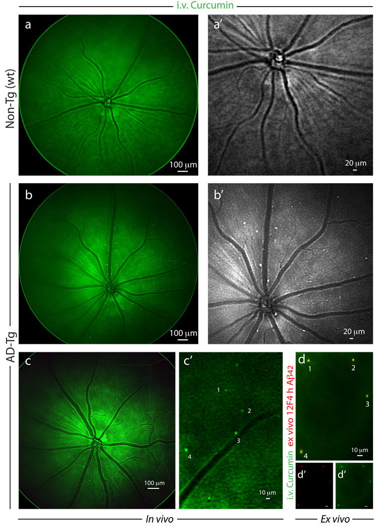
Noninvasive in vivo optical imaging of curcumin-labeled Aβ plaques in AD-Tg mice retina. (a–c) Following i.v. administration of curcumin (7.5 mg/kg) for 5 consecutive days, retinas of 8–12 month-old AD-Tg and wt mice were in vivo fluorescently imaged using Micron II rodent retinal microscope. Representative in vivo mouse fundus images: (a) no plaques could be visualized in wt mouse; (a’) higher magnification in grayscale. (b) Retinal curcumin-labeled plaques (green spots) were visible in live AD-Tg mouse; (b’) higher magnification in grayscale. (c,c’) Representative images demonstrate in vivo detection of curcumin-labeled plaques in the retinas of 8 month-old AD-Tg mice, following a single i.v. injection of curcumin (7.5 mg/kg) two hours prior to imaging. (c’) Enlarged image of the selected area from (c) image, demonstrates that this imaging modality enables the identification of individual plaques or plaque clusters at a high spatial resolution. Note that blood vessels appear unstained (dark), possibly due to blood flow in the live mouse. (d) Whole-mount retina prepared from the same in vivo imaged mouse eye as in (c), following perfusion. An additional ex vivo staining with anti-human Aβ42 mAb (12F4; secondary Ab-Cy5 conjugate) further confirmed the specificity of curcumin signals (captured by Micron II) to Aβ plaques. (d’,d”) separate channels for each staining. A similar pattern of curcumin-positive plaques imaged in vivo was identified ex vivo following antibody labeling (indicated by the numbers 1–4).

