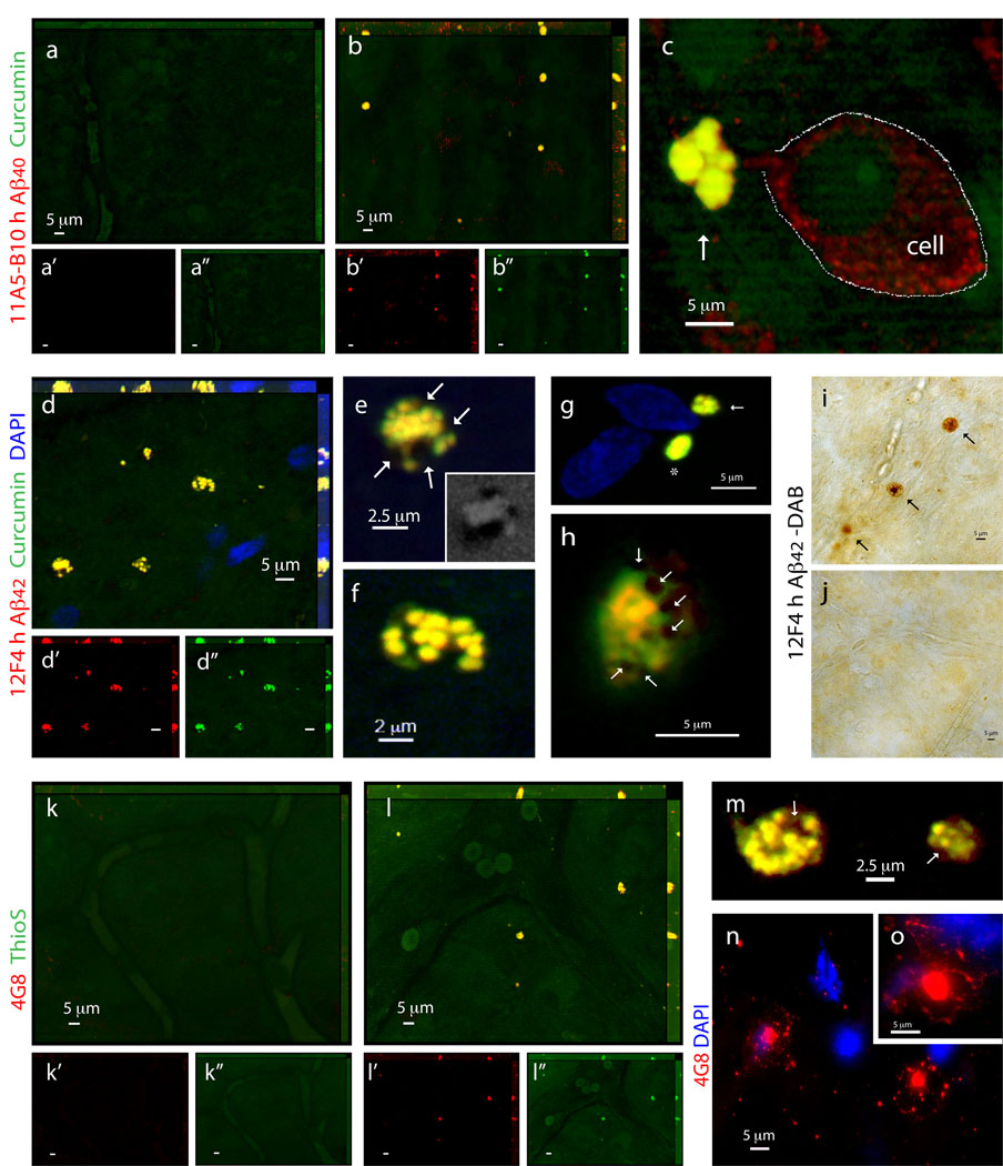
Figure 6.
Characterization of retinal Aβ plaques identified in postmortem retinas of definite AD patients. (a–c) Representative z-axis projection images of whole-mount retinas of (a) normal individuals compared to (b,c) AD patients following curcumin and anti-human Aβ40 mAb 11A5-B10 stainings. (a) No Aβ plaques could be detected in normal control retinas, whereas (b) clearly found in retinas from AD patients. (a’–b”) Separate channels for each staining. (c) At higher magnification, extracellular Aβ plaque with compacted large cluster is indicated by an arrow (intracellular Aβ40 is demarcated by a dotted line). (d–h) Whole-mount retinas from AD patients stained with curcumin and anti-human Aβ42 mAb 12F4. (d’,d”) Separate channels. Note co-localization of curcumin and antibody. (e,f) Higher magnification images of Aβ plaques demonstrated their compacted morphology, consisting of multiple small dense cores connected in larger cluster. (e) Aβ plaques containing lipid deposits indicated by arrows; right bottom image captured in DAPI channel shows dark spots of SBB staining representing lipid deposits associated with retinal Aβ plaque. (g) Aβ plaques stained with curcumin and 12F4 mAb, display either compacted single-globular (asterisk) or cluster (arrow) morphology, both lack notable lipid-associated deposits. (h) Aβ plaque with compacted morphology and associated dark SBB staining spots suggesting the presence of lipid deposits (arrows). (i,j) Immunoperoxidase staining of Aβ plaques labeled with primary mAb 12F4 (plaques are indicated by black arrows) in retinal whole-mount from (i) AD patient and (j) non-AD control. DAB was used as a chromogen. (k–m) Representative retinas from (k) normal individual compared to (l) AD patient stained with ThioS and anti-Aβ mAb 4G8. ThioS- and 4G8 double-positive parenchymal Aβ plaques are found in AD patients’ retinas but not in normal controls. (k’–l”) Separate channels. (m) Higher magnification demonstrating compacted morphology of Thio-S-positive retinal Aβ plaques in AD patients’ retinas. (n,o) Single-labeled Aβ plaques using 4G8 mAb (secondary Ab-Cy5 conjugate) in the retinal innermost layers: Aβ plaques have classical morphology consisting of a central dense-core and radiating fibrillar arms.

