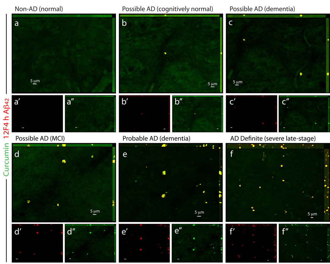
Figure 7.
Detection of retinal Aβ plaques in suspected early AD patients. (a–f) Representative whole-mount retinas from normal individuals (Non-AD) and from possible/probable AD patients (based on the combined clinical diagnosis and postmortem brain pathology; Table S1), stained with curcumin and anti-Aβ42 mAb (12F4; secondary Ab-Cy5 conjugate). (a) In the retina from cognitively normal individual with no AD pathology, Aβ plaques could not be detected. (b) In cognitively normal individual with mild Aβ-plaque brain pathology (mainly diffused plaques), sparse retinal Aβ plaques were identified. (c,d) In patients with possible AD diagnosis, clusters of Aβ plaques were observed, and (e) in retinas from patients with several years of dementia and postmortem diagnosis of probable AD, Aβ plaques were more common throughout the retina. (f) Abundant Aβ plaques were observed in the retina from severe-stage AD definite patient (based on combined clinical diagnosis and postmortem brain pathology; Table S1). (a’–f”) separate channels for each staining.

