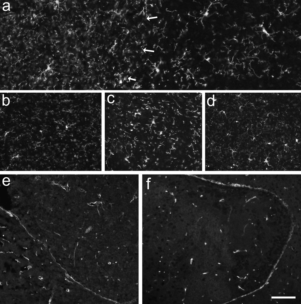
Figure 4.
The pan-microglia marker Iba1-immunoreactivity (a–d) demonstrated that the density of microglia was slightly higher in the striatal portion of co-grafts (a; left to arrows) compared to the VM (a; right). There was no increase in microglia over time as found at 12 months postgrafting in Gdnf +/+ (a) compared to Gdnf +/− (b) transplants, although the Gdnf +/− transplants had a significant loss of TH-positive neurons at this time point. Comparing the VM portion of co-grafts, no difference between Iba1-positive microglia was demonstrated between Gdnf +/+ (c) and Gdnf −/−(d) at 3 months postgrafting. Using Glut1-immunohistochemistry to study blood vessel support in the transplants demonstrated that both the Gdnf+/+ (e) and Gdnf−/−(f) transplants had good and evenly distributed blood vessel support. Scale bar: a–d =50 µm, e, f = 100 µm.

