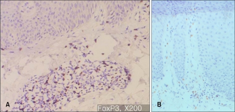
Fig. 2.
(A) Immunohistochemistry for FOXP3, ×200: Typical plaque type psoriasis (Case No. 3). In psoriatic skin lesions, FOXP3+ cells are mainly observed in papillary dermis, consisting of about 20~40% of the mononuclear cellular infiltration. (B) Immunohistochemistry for FOXP3, ×200: Palmoplantar pustular psoriasis (Case No. 15). FOXP3+ cells consists of about 20~30% of the mononuclear cellular infiltration.

