
Figure 1.
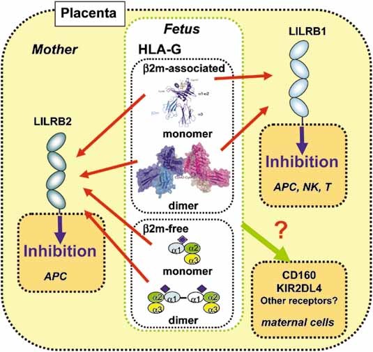
Receptor recognition of the different forms of HLA-G at the maternal-fetal interface. HLA-G can be expressed in various forms in placenta. The structures of β2m-associated HLA-G monomer and dimer are shown. LILRB1 (light blue), which is expressed on decidual antigen-presenting cells (APC) and subsets of NK and T cells, binds to only β2m-associated forms of HLA-G monomer and dimer. LILRB2 (dark blue), whose expression is restricted to decidual APC, binds to both β2m-associated and -free forms of HLA-G monomer and dimer. LILRB1 and LILRB2 presumably bind to HLA-G dimer more strongly than its monomer. The thickness of the arrows corresponds to the signal strength. Both membrane-bound and soluble forms likely inhibit the immune responses of LILRB-expressing cells. On the other hand, the HLA-G recognition of CD160 and KIR2DL4 still remains unclear.