
Figure 2.
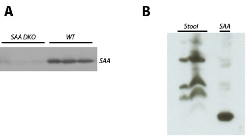
SAA levels in plasma and stool of knockout and wildtype mice. Samples of acute phase plasma, obtained 24 h after intravenous injection of 1 microgram LPS into wildtype mice or DKO mice (A), or extracts of stool samples from wildtype mice (B) were analyzed by immunoblotting with anti SAA1/2 antiserum following SDS-PAGE. The right lane in "B", labeled "SAA", is a positive control consisting of acute-phase plasma obtained as described for panel A. The reduced mobility of SAA in stool samples is likely caused by self-aggregation of SAA.