
Fig. 4.
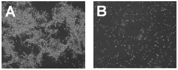
Phase contrast microscopy images illustrating the morphology of C. albicans ATCC 10231 cultures grown with vehicle only (A) or treated with compound 1 (B). Whereas the mycelia morphology of the control C. albicans cells appeared as mats of tangled filaments, mycelia were not apparent in cells treated with 1. For the experiment, overnight shake cultures of C. albicans were prepared under aerobic conditions in yeast extract-peptone medium with 2% glucose. The cells were diluted in fresh medium (1 : 100) and 100 μL aliquots added to the wells of a flat-bottom 96-well plate. Cells were treated with DMSO only (vehicle control) or compound 1 (2 μL of compound prepared in DMSO at 5 mg mL−1).