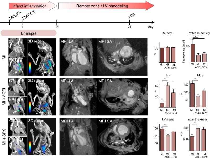
Figure 7. Enalapril reduces early protease activity and subsequently LV remodeling in apoE-/- mice with MI.
Top: Flow chart illustrating the set up of the study.
Left: FMT-CT 1 day after MI. Arrows denote apical infarct signal. Additional activation of the protease sensor is seen in the vicinity of the decending aorta and may be due to inflammatory atherosclerotic lesions. Infarct size was measured by contrast-enhanced CT.
Middle and right panels: Cardiac MRI, LA: long axis, SA: short axis view. ACEi: Enalapril treatment, SPX: splenectomy. Mean ± SEM; * p < 0.05.

