FIGURE 1.
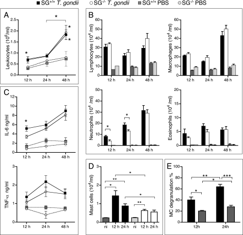
Accumulation of leukocytes in T. gondii-infected SG+/+ and SG−/− mice. Peritoneal exudate cells were collected 12, 24, and 48 h after intraperitoneal inoculation of 106 RH strain tachyzoites or PBS and analyzed as described under “Experimental Procedures.” A, leukocyte recruitment into exudate fluids after infection of SG+/+ and SG−/− mice. B, differential counting of peritoneal leukocytes on cytospin slides stained with May-Grünwald Giemsa. C, levels of the proinflammatory cytokines IL-6 and TNF-α in peritoneal exudates were determined by ELISA. D, MCs were analyzed by counting CD117+ cells by flow cytometry. Samples from noninfected (ni) mice were used as controls. E, the proportion of MCs that were degranulated in infected and naïve SG+/+ mice were determined. Results are expressed as means ± S.E. Pooled results from at least two independent experiments are shown with n = 7, 10, and 12 for SG−/− mice, and n = 6, 10, and 11 for SG+/+ mice at 12, 24, and 48 h, respectively. *, p ≤ 0.05; **, p < 0.001; ***, p < 0.0001.

