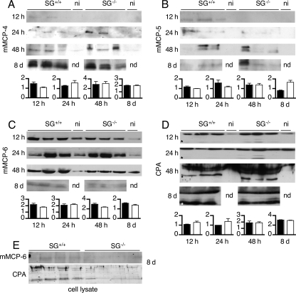
FIGURE 3.
Peritoneal challenge with T. gondii leads to MC protease secretion in SG-deficient animals. Cell-free peritoneal lavage fluid was collected from T. gondii-infected and noninfected (ni) animals at 12, 24, and 48 h and 8 days post-infection. The lavage fluids were concentrated, and equal amounts of protein were separated by SDS-PAGE and transferred to membranes. Western blots were developed with antibodies against the respective enzymes, and for each time point, the mean signal intensity minus background for the proteases is given as an inset graph as described under “Experimental Procedures.” Black bars represent SG+/+, and open bars represent SG−/−. Representative blots for mMCP-4 (A), mMCP-5 (B), mMCP-6 (C), and MC-CPA (D) from infected (n = 3) and noninfected (n = 1) SG+/+ and SG−/− mice are shown. E, Western blots for mMCP-6 and MC-CPA of cellular lysates 8 days post-infection. Asterisks indicate the expected position of the active form of MC-CPA. nd, not determined.

