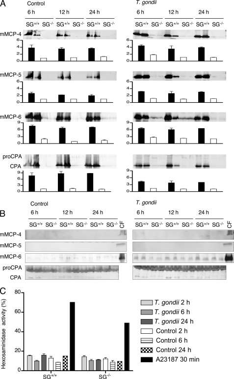
FIGURE 4.
In vitro stimulation of peritoneal derived mast cells with live T. gondii. A, SG+/+ and SG−/− PCMCs (106 cells) were stimulated with 106 live T. gondii tachyzoites for the indicated time periods. Nonstimulated (control) and stimulated cells were then analyzed by Western blotting for the indicated MC proteases. Western blots were developed with antibodies against the respective enzymes, and for each time point, the mean signal intensity minus background for the proteases is given as an inset graph as described under “Experimental Procedures.” Black bars represent SG+/+, and open bars represent SG−/−. In B, representative Western blots of supernatants from T. gondii-stimulated or control PCMCs are shown. The two bands shown for each time point and genotype represent two independent stimulations performed in separate wells. Cell fractions (CF) of SG+/+ PCMCs were included as positive controls (C). As a control of degranulation, β-hexosaminidase release was measured from PCMCs after stimulation with T. gondii for 2, 6, and 24 h. The calcium ionophore A23187 was included as a positive control for degranulation. Results are presented as means of three animals of each genotype ± S.E.

