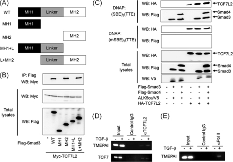
FIGURE 6.
TGF-β-dependent interaction of TCF7L2 with TTE. A, depiction of Smad3 mutants. MH, mad homology region. B, interaction of Smad3 with TCF7L2 via its MH2 domain. Immunoprecipitations were carried out using anti-FLAG M5 antibody, and coimmunoprecipitated (IP) TCF7L2 was detected by Western blotting (WB) using anti-Myc 9E10 antibody (top panel). The expression of Myc-TCF7L2 and Smad3 mutants conjugated with FLAG at the N terminus was evaluated using anti-Myc 9E10 (middle panel) and anti-FLAG M5 antibodies (bottom panel), respectively. C, requirement of Smad-SBE complex for enhanced binding of TCF7L2 to TTE. COS7 cell lysates transfected with the plasmids indicated were mixed with either biotinated (SBE)3(TTE) or biotinated (mSBE)3(TTE). TCF7L2-DNA complex and Smads-DNA complex were detected by Western blotting using anti-HA3F10 antibody (top and third panels) and anti-FLAG M5 antibody (second and fourth panels), respectively. The expressions of HA-TCF7L2, FLAG-Smads, and ALK5ca/V5 were evaluated using anti-HA3F10 (fifth panel), anti-FLAG M5 (sixth panel), and anti-V5 antibodies (bottom panel), respectively. D and E, recruitment of TCF7L2 to the C region including TTE upon TGF-β stimulation. Cross-linked chromatin from HepG2 cells were incubated with anti-TCF7L2 (D) and anti-RNA polymerase II antibodies (E). The immunoprecipitated DNA was analyzed by PCR with primers that amplify the fragment including TTE in the TMEPAI gene. As a positive control, DNA sequences including TBE in the TCF7 gene were also amplified (lower panel in D). As a negative control, PCR was performed using DNA immunoprecipitated with mouse control IgG. In parallel, the input DNA was amplified by PCR as described above.

