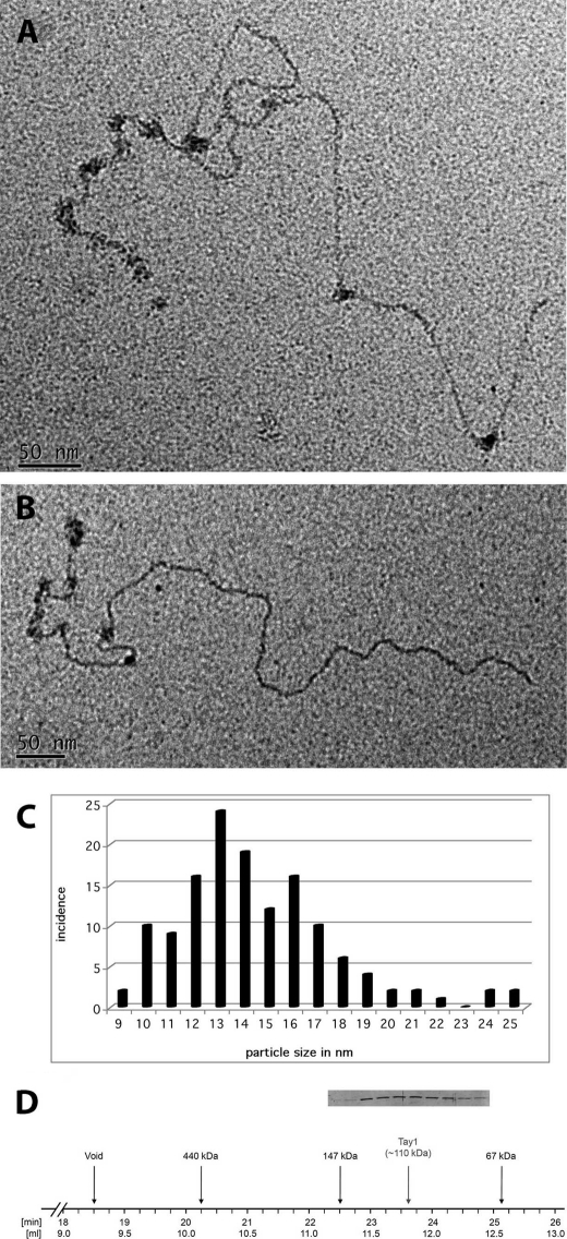
FIGURE 6.
Estimation of the size of the Tay1p particles bound to DNA by positive staining. Complexes of Tay1p on the model Y. lipolytica telomere DNA were formed as described in Fig. 4 (see also “Experimental Procedures”) and prepared for EM by adsorption to thin carbon foils and staining with 2% uranyl acetate. A and B, Tay1p binds preferentially along the 810-bp telomeric tract but occasionally elsewhere on the DNA. Measurement of the diameter of 127 particles from examples such those shown in A and B generated a size distribution shown in C. D, when analyzed by gel filtration on Superdex-200, the peak of Tay1p is eluted in the fractions corresponding to dimeric forms of the protein. The lanes in the inset correspond to the fractions indicated on the horizontal line.

