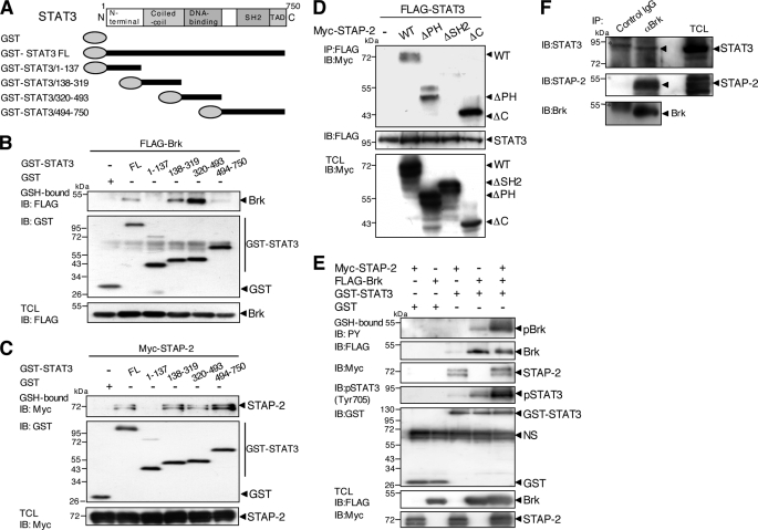
FIGURE 7.
Molecular interactions among STAP-2, STAT3, and Brk. A, schematic diagrams of the domain structures of STAT3 and a deletion mutant are shown. B, 293T cells (1 × 10⁷) were transfected with FLAG-tagged Brk (10 μg) with or without GST-fused STAT3 FL or a deletion mutant (10 μg). At 48 h after transfection, the cells were lysed, pulled down with glutathione-Sepharose, and immunoblotted (IB) with an anti-FLAG or anti-GST antibody. An aliquot of each TCL was immunoblotted with an anti-FLAG antibody. C, 293T cells (1 × 10⁷) were transfected with Myc-tagged STAP-2 (10 μg) with or without GST-fused STAT3 FL or a deletion mutant (10 μg). At 48 h after transfection, the cells were lysed, pulled down with glutathione-Sepharose, and immunoblotted with an anti-Myc or anti-GST antibody. An aliquot of each TCL was immunoblotted with an anti-Myc antibody. D, 293T cells (1 × 10⁷) were transfected with FLAG-tagged STAT3 (10 μg) with or without Myc-tagged STAP-2 deletion mutants (8 μg). At 48 h after transfection, the cells were lysed, immunoprecipitated (IP) with an anti-FLAG antibody, and immunoblotted with an anti-Myc or anti-FLAG antibody. An aliquot of each TCL was immunoblotted with an anti-Myc antibody. E, 293T cells (1 × 10⁷) were transfected with or without FLAG-tagged Brk (10 μg) with or without GST-STAT3 FL (10 μg) and/or Myc-tagged STAP-2 (8 μg). At 48 h after transfection, the cells were lysed, pulled down with glutathione-Sepharose, and immunoblotted with an anti-PY, anti-Myc, anti-pSTAT3 (Tyr-705), anti-FLAG, or anti-GST antibody. An aliquot of each TCL was immunoblotted with an anti-FLAG or anti-Myc antibody. F, human breast cancer T47D cells (3 × 10⁷) were lysed, immunoprecipitated with control IgG or anti-Brk antibody, and immunoblotted with anti-STAT3, anti-STAP-2, or anti-Brk antibody. The asterisk shows a nonspecific band.

