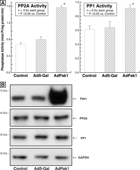
FIGURE 3.
Cardiomyocyte PP2A and PP1 activity and quantity after AdPak1 infection. The feline cardiomyocytes used for these assays were either uninfected (control) or infected with AdPak1 or Adβ-gal 72 h earlier. A, PP2A activity was measured using an immunoprecipitation assay kit (catalog number 17-313; Upstate Biotech) as described under “Experimental Procedures.” PP1 activity was determined using the PSP assay system (catalog number P0780S; New England Biolabs), as is also described under “Experimental Procedures.” B, these immunoblots were prepared from the same cardiomyocyte lysates used for the PP2A and PP1 activity assays. The blots were prepared using a polyclonal anti-Pak1 (α-Pak) antibody (sc-881; Santa Cruz Biotechnology), a monoclonal antibody to the catalytic subunit of PP2A (clone 1D6; Upstate Biotech), a monoclonal antibody to PP1 (clone E-9; Santa Cruz Biotechnology), and, as the loading control, a monoclonal antibody to GAPDH (clone 6C5; Upstate Biotech). *, p < 0.05 by one-way ANOVA with Bonferroni post hoc analysis.

