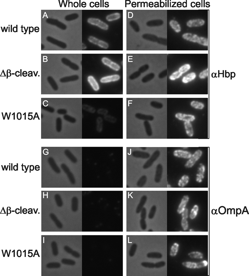
FIGURE 6.

HbpW1015A cannot be detected at the cell surface by immunofluorescence. MC1061degP::S210A cells expressing Hbp or the indicated mutant Hbp derivatives or carrying an empty vector (pEH3) were collected 1 h after induction with 200 μm IPTG and subjected to indirect immunofluorescence using a polyclonal antiserum against either the Hbp passenger or OmpA and subsequently a Cy3-labeled conjugate (right panels). In addition, the corresponding fields are shown by phase-contrast microscopy (left panels). The OM of half of the cells was permeabilized prior to labeling, as indicated and described under “Experimental Procedures.” Δβ-cleav., Δβ-cleavage.
