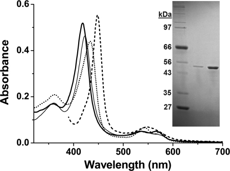
FIGURE 1.
Purification and spectral features of CYP142. Spectra in the main figure show the UV-visible absorption features of pure, substrate-free ferric M. tuberculosis CYP142 (3.7 μm) (thick solid line) and for CYP142 bound to econazole (13 μm, thin solid line), nitric oxide (dotted line), and in the Fe2+-CO form bound to cholest-4-en-3-one (10 μm, dashed line). The major (Soret) absorption band is centered at 418, 423, 433.5, and 448 nm, respectively. The inset shows an SDS-polyacrylamide gel with molecular weight markers of indicated mass in the 1st lane and purified CYP142 (0.5 and 2.5 μg) as a single band in the remaining lanes.

