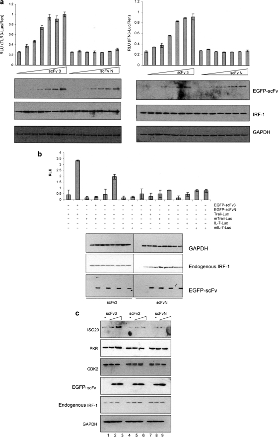
FIGURE 6.
scFv activates endogenous IRF-1. a, HeLa cells were transfected with a titration of EGFP-scFv3 or EGFP-scFvN (0–250 ng) plus Renilla (60 ng) and either TLR3-Luc (120 ng) (left-hand graph) or IFNβ-p125luc (right-hand graph) (120 ng). DNA levels were normalized using an empty vector. Reporter gene activity was measured in relative light units (RLU) and is expressed as the ratio of Luc/Ren. The assays were carried out in duplicate; results are given as mean ± half the range and are representative of at least three independent sets of experiments. Expressed proteins were detected by immunoblot using anti-EGFP and anti-IRF-1, and GAPDH was detected as a loading control. b, HeLa cells were transfected with either TRAIL-Luc (WT or mutant-ISRE) or IL-7-Luc (WT or mutant -ISRE) (120 ng), in each case with or without EGFP-scFv3 or EGFP-scFvN (120 ng). Reporter assays and immunoblots were carried out as above. c, lysate from HeLa cells transfected with 0, 0.5, or 1 μg of EGFP-scFv3 (lanes 1–3), EGFP-scFv2 (lanes 4–6), or EGFP-scFvN (lanes 7–9) were analyzed by SDS-PAGE/immunoblot and probed with anti-ISG20, anti-PKR, anti-CDK2, anti-EGFP, anti-IRF-1, or anti-GAPDH antibody.

