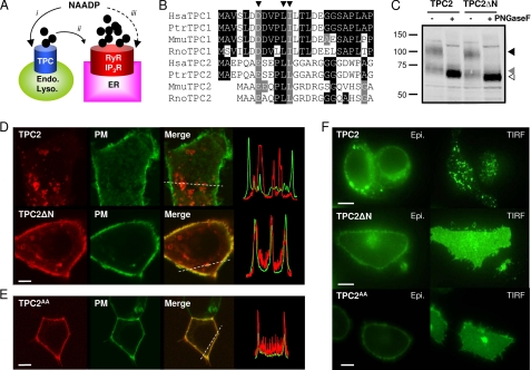
FIGURE 1.
Removal of the N terminus redirects TPC2 from lysosomes to the plasma membrane. A, possible actions of NAADP. NAADP is proposed to activate TPCs on the endo-lysosomal system (i) followed by amplification of these trigger signals by ER Ca2+ channels (ii) such as ryanodine receptors (RyR) and inositol trisphosphate receptors (IP3R). Alternatively, NAADP has been proposed to activate Ca2+ release from the ER directly (iii). B, sequence alignments of Human (Hsa), chimpanzee (Ptr), mouse (Mmu) and rat (Rno) TPC N-terminal sequences highlighting a conserved dileucine endo-lysosomal targeting motif conforming to the consensus sequence (DE)XXXL(L/I) (arrowheads). C, Western blot analysis of SKBR3 cell extracts expressing GFP-tagged TPC2 or TPC2ΔN (10 μg/lane). Samples were incubated with (+) or without (−) peptide:N-glycosidase F (PNGaseF) prior to analysis. Arrowheads mark the positions of fully glycosylated (black arrowhead), core glycosylated (gray arrowhead), and deglycosylated (white arrowhead) TPC2 or TPC2ΔN. Results are representative of three experiments. D, confocal fluorescence images of SKBR3 cells coexpressing GFP-tagged human reduced folate carrier to delineate the plasma membrane (PM) and either mRFP-tagged TPC2 (top) or mRFP-tagged TPC2 lacking residues 3–25 (TPC2ΔN; bottom). Merged images are overlays of the plasma membrane marker (green) and TPC constructs (red). Transect analyses of red and green fluorescence intensities across the regions marked by dotted lines are shown on the right. E, confocal fluorescence images of HEK cells coexpressing the plasma membrane marker and mRFP-tagged TPC2 in which Leu-11 and Leu-12 were replaced with Ala (TPC2AA). F, epifluorescence (Epi.) and TIRF images of HEK cells expressing C-terminally GFP-tagged TPC2, TPC2ΔN, or TPC2AA. Images (D–F) are typical examples from samples of 15–20 cells. All scale bars, 5 μm.

