FIGURE 5.
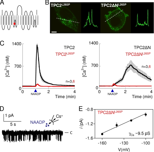
TPC2 is the pore-forming subunit of an NAADP-gated channel. A, depiction of TPC2 showing location of a putative pore residue (Leu-265, red). B, confocal fluorescence images of SKBR3 cells expressing GFP-tagged TPC2 in which Leu-265 was replaced by proline (TPC2L265P, left) or in which this was combined with removal of the N terminus (TPC2ΔNL265P, right). Images are typical of those from 6–10 cells. Scale bar, 5 μm. Similar results with HEK cells are shown in supplemental Fig. S2E. C, cytosolic Ca2+ signals from individual fura-2-loaded SKBR3 cells transiently transfected with the indicated C-terminally GFP-tagged TPC2 constructs and microinjected with NAADP (10 nm, arrowheads). Results are means ± S.E. of the indicated number (n) of cells. D, recording, typical of four similar records, from excised inside-out patches from the plasma membrane of HEK cells expressing TPC2ΔNL265P and stimulated with 500 nm NAADP in the bathing solution with Cs+ as the charge carrier. C denotes the closed state. E, current-voltage relationship from records similar to those shown in D. Results are means ± S.E., n = 4.

