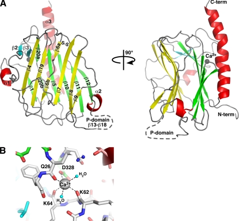
FIGURE 1.
Structure of the CRT lectin domain. A, schematic representation of the structure. Helices are shown in red, β-strands in the concave β-sheet are yellow, the convex β-sheet is green, and the two additional strands β2 and β3 are cyan. The bound calcium ion is shown as a gray sphere. The position of the P-domain that contains strands β13–β20 is indicated by a dashed line. Cys105 and Cys137 in the concave β-sheet form a disulfide bond (S–S). B, enlarged view of the calcium-binding site. Residues and hydrogen bonds (dashed lines) coordinating the calcium ion are shown along with two coordinating water molecules (cyan spheres). Residue color coding is the same as in A. N-term, N terminus; C-term, C terminus.

