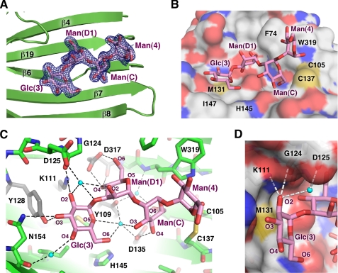
FIGURE 2.
Structural basis of Glc1Man3 recognition by CRT. A, omit map calculated in the absence of tetrasaccharide shows well defined electron density (blue) for all four sugar moieties. The tetrasaccharide binds in a cavity on the concave β-sheet. B, surface representation of CRT shows the side chains of Phe74, Met131, His145, Ile147, Trp319, and the Cys105–Cys137 disulfide bridge form the walls of the cavity in contact with the glycan (magenta). C, oxygens in the tetrasaccharide form a network of hydrogen bonds (dotted lines) with ordered water molecules (cyan spheres) and CRT. Residues that disrupt CRT binding when mutated are shown in gray (17–19). D, the equatorial oxygen (O2) of glucose makes hydrogen bonds with the side chain of Lys111 and backbone carbonyl of Gly124. Mannose has an axial O2, which clashes sterically with the underlying side chain of Met131 to prevent binding in that position.

