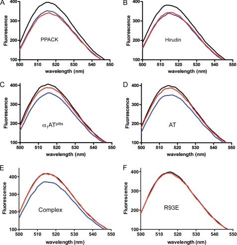
FIGURE 1.
The fluorescence spectra of fluorescein-labeled fibrinogen γ′-peptide in the presence of free and inhibited thrombin. In each case, the black trace is the solution containing fluorescein-γ′ alone, the blue trace is after the addition of thrombin to the solution, and the red trace is after the further addition of a thrombin inhibitor (A, PPACK; B, hirudin; C, α1ATpitts; and, D, AT). E, no fluorescence quench is observed when thrombin inhibition is complete. The black trace is buffer with the fluorescein-γ′ alone; red is after the addition of preformed α1ATpitts-thrombin complex; and blue is fluorescein-γ′ with uninhibited thrombin. F, exosite II variant R93E is deficient in fluorescein-γ′ binding, although a small quench is observed upon the addition of the thrombin variant (red).

