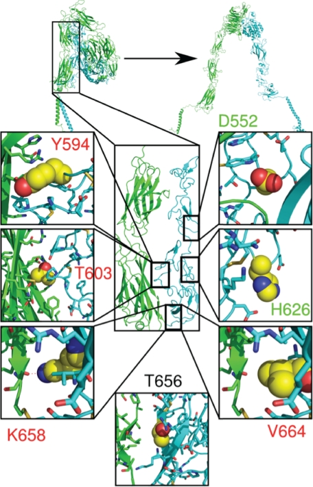
FIGURE 3.
Location of the designed β3 mutations in the interface of the αIIb and β3 stalks. The seven mutated β3 residues are shown in the crystal structure reported by Zhu et al. (4). The αIIb stalk is shown in green and the β3 stalk in cyan. The boxes in the figure show the side chains of the mutated WT residues as yellow spheres. Alanine mutations predicted to be destabilizing and neutral are lettered in red and green. A residue predicted to be stabilizing when mutated to tryptophan is indicated by black lettering.

