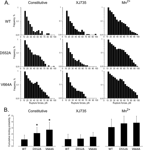
FIGURE 5.
Effect of β3 stalk mutations on OPN binding to αvβ3 as measured by laser tweezers-based rupture force spectroscopy. Rupture forces between latex beads coated with OPN and CHO cells expressing WT αvβ3 or αvβ3 containing the β3 mutations D552A and V664A were measured as described under “Experimental Procedures.” A, histograms showing the distribution of rupture forces between the OPN-coated beads and αvβ3-expressing CHO cells in the absence of an αvβ3 activator (Constitutive), in the presence of the αvβ3-specific antagonist 100 μm XJ735, and in the presence of the αvβ3 activator 1 mm Mn2+. Measured rupture forces were sorted into histograms and normalized by the total number of bead-cell contacts. The percentage of events in a particular force range (bin) represents the percent frequency of a bond rupture in that range. B, cumulative probabilities of detecting rupture forces >10 pN, normalized by the level of αvβ3 expression, between OPN-coated beads and αvβ3-expressing CHO cells under the conditions described in A. Data shown are the mean ± S.D. (error bars) of 15–40 individual experimental files. *, t tests for statistical significance: D552A versus WT, p = 0.02043; V664A versus WT, p = 0.0076.

