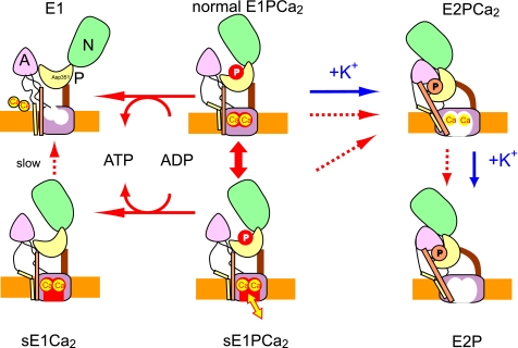
FIGURE 7.
Schematic model for roles of K+ in EP processing and Ca2+ handling in Ca2+ transport. sE1PCa2 is an E1PCa2 species formed without K+ possessing a closed cytoplasmic gate and lumen-facing Ca2+ binding sites (an opened lumenal pathway) with high Ca2+ affinity (Fig. 6). sE1PCa2 is in rapid equilibrium with the normal E1PCa2. Here, s denotes silent because this species is apparently absent in the presence of K+ and also because the bound Ca2+ ions are not released to the cytoplasmic side even upon ADP-induced reverse dephosphorylation (to sE1Ca2) in contrast to the normal E1PCa2 reverse dephosphorylation. Actual active Ca2+ transport is achieved by a large reduction of the Ca2+ affinity during the normal sequence E1PCa2 → E2PCa2 → E2P + 2Ca2+ (blue arrows). The schematic is based on crystal structural models for the ADP-sensitive and -insensitive EP states and E1Ca2, with the positions of the cytoplasmic N, P, and A domains, and membrane (orange layer) being approximate. The Ca2+ sites in the transmembrane domain are depicted as occluded (closed cytoplasmic and lumenal gates) in normal E1PCa2, as lumen-facing and high Ca2+ affinity with the closed cytoplasmic gate in sE1PCa2 and sE1Ca2, and as lumenally opened with reduced Ca2+ affinity in E2P and E2PCa2 (immediately before the Ca2+ release).

