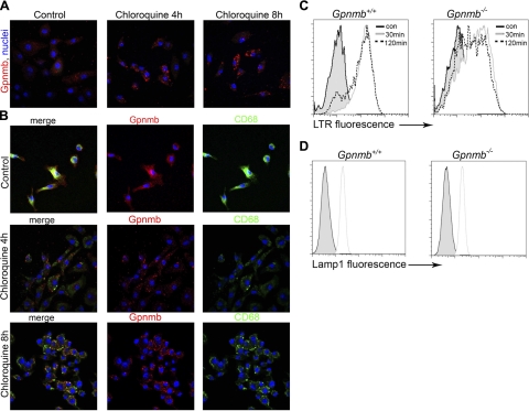
Figure 7.
Characterization of intracellular compartments in Mφs. A) Confocal images of d 7 BMMφs, untreated or treated with chloroquine, then immunolabeled for Gpnmb. B) Split-panel confocal images of CD68 and Gpnmb in BMMφs untreated or treated with chloroquine. C, D) Flow cytometric histogram plots of LysoTracker Red fluorescence (C) and LAMP-1 immunofluorescnce (D) in WT and Gpnmb mutant BMMφs after 30 or 120 min loading. Note a population of LTR dim cells in Gpnmb mutants.

