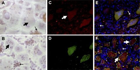
Figure 6.
Immunostaining for ANGPTL4. A, B) In vitro differentiated osteoclasts cultured on dentine slices were exposed to hypoxia (2% O2, 24 h) and stained with anti-ANGPTL4 (A) or anti-HIF-1α (B). C–F) Immunofluorescence of GCTB sections stained for ANGPTL4 (C), HIF-1α (D), and merge: DAPI (blue), ANGPTL4 (red), and HIF-1α (green) (E) and DAPI (blue), ANGPTL4 (red), and VNR (green) (F). Broad arrows indicate osteoclasts; narrow arrows indicate mononuclear cells.

