
Fig. 1.
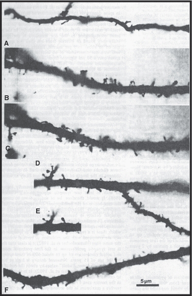
Micrographs of segments of apical dendrites of layer III cortical pyramidal neurons. (A) Fetus at 33 weeks' gestation. Narrow dendrite with few immature, hair-like spines. (B,C) Thicker dendrite at 3 months postnatal with more bulbous spines. (D,E) Spine numerical density is highest at 5 months. (F) 21 months: spine density has fallen to perinatal levels. From Michel & Garey (1984).