
Fig. 2.
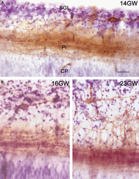
Morphology of human CR cells and their axonal plexus, visualized with DiI tracing followed by photoconversion and Nissl-staining. (A) At 14 GW, CR cells within the subpial granular layer (SGL) extend the dense axonal plexus (Pl) at the interface of marginal zone (MZ) and cortical plate (CP). (B) At 16 GW, CR cells change to a deeper position in the MZ, below the SGL. (C) At 23 GW. CR cells display bizarre morphologies, whereas the SGL cells disperse throughout the MZ. Scale in (A) for A–C: 20 μm.