
Fig. 3.
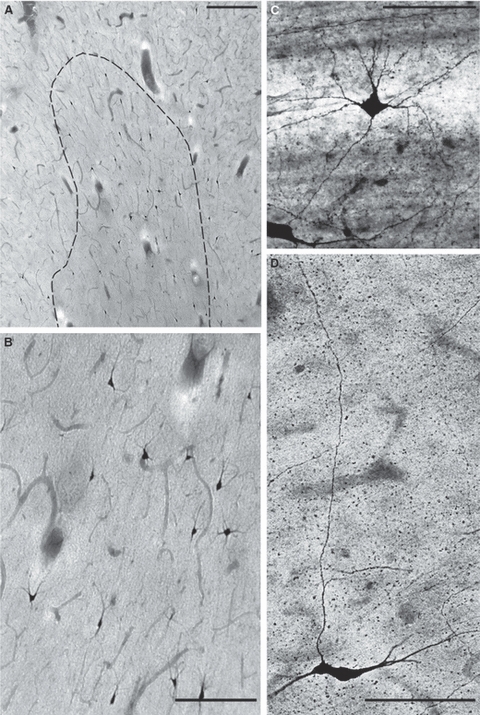
Nitrinergic (NADPH-diaphorase-stained) interstitial neurons in the gyral white matter (right inferior frontal gyrus) at 3 years (A,B; B is enlarged part of A) and 12 years (C,D). Note the multipolar nitrinergic interstitial neuron closely apposed to the blood vessel (C) and fusiform interstitial neuron, which sends a long ascending axon to the overlying cortex (D). Scale bars: 150 μm (A,B) and 100 μm (C,D).