
Fig. 2.
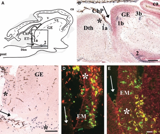
(A) Lateral sagittal section through a 5.5-gestational week (gw) human embryo showing ionized calcium-binding adapter molecule 1 (Iba1)-positive cells issued from the choroid plexus (ChP) and constituting patch 1a in the eminentia thalami (EM/ET). Patch 2 is contiguous to the ditelencephalic fissure (arrows). Grey areas vary in colour depending on the Iba1-positive cell density. The box corresponds to C, where patches of microglial cells are indicated by asterisks. (B,C) Iba1 (black)-CD34 (brown) double labelling and counterstaining with neutral red. (B) Sagittal section through an 8-gw embryo [level indicated in Fig. 2G in Monier et al. (2006)]. Iba1-positive patches 1a, 1b, and 2 are visible near the ditelencephalic fissure (arrow), patch 1a near the presumptive anlage of the internal capsule, and patch 3b at the presumptive anlage of the external capsule. (D,E) High magnification at the level of ChP and ET shown in A. Fluorescent double labelling for Iba1 (green) and CD45 (red) (D) and for Iba1 (green) and CD68 (red) (E) showing co-localization (yellow). Note the preferential CD68 labelling deeper in the EM parenchyma (asterisk in E) compared with the preponderant CD45 labelling in the ChP (asterisk in D). ca, cortical anlagen; Dien, diencephalon; GE, ganglionic eminence; post, posterior; Dth, dorsal thalamus; VZ, ventricular zone; V, ventricle. Scale bars: B, 500 μm; C, 100 μm; D and E, 25 μm. Adapted from Figs 1 and 2 in Monier et al. (2006).