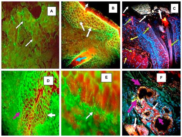
Fig 2.
A, B, D, and E, Results of indirect IF (IF) performed after partial fixation with paraformaldehyde. C and F, Results of indirect immunofluorescence performed using paraffin-fixed samples followed by antigen retrieval techniques. A, Using conjuctiva as the antigen source, antihuman conjugated IgG-Alexa 488 (Invitrogen) showed positive ICS and basement membrane zone (BMZ) staining (white arrows). B, Positive intercellular (IC), BMZ, and hair follicle staining using antihuman total IgG antiserum as secondary antibody (green) (fluorescein isothiocyanate [FITC]) (white arrows). Nuclei of cells were stained with Topro III (Invitrogen) (red ). In addition, note strong reactivity of hair follicle bulb to Ki-67 proliferating antigen (red) (Texas Red) (blue arrow). C, Structure (top) that resembles secretion near tear duct, likely of mixed material including mucins, lipids, and other tear components. These components contribute to high, non-Newtonian viscosity of tear film and its low surface tension, features essential for tear film stability (white arrows). In same figure, Ki-67 antigen demonstrated clumped elongated pattern around eyelid base, within isthmus, and in some parts of epidermal layer (red) (green arrows). Positive autoreactivity as small and large dots (red) using Alexa Fluor 555 (Invitrogen) against human IgG (yellow arrows). Nuclei were counterstained with DAPI (Pierce) (blue). D and F, IC and BMZ staining were seen in El Bagre-endemic pemphigus foliaceus using antihuman conjugated IgG FITC (white arrows)(×20). D, BMZ staining of meibomian glands (×100). E, Secretory portion of meibomian gland, in yellow dots, as part of intrinsic fluorescence of these structures (purple arrows). Ki-67 antigen showed positive clumped pattern surrounding involving base of gland ducts on eyelid (white arrows).

