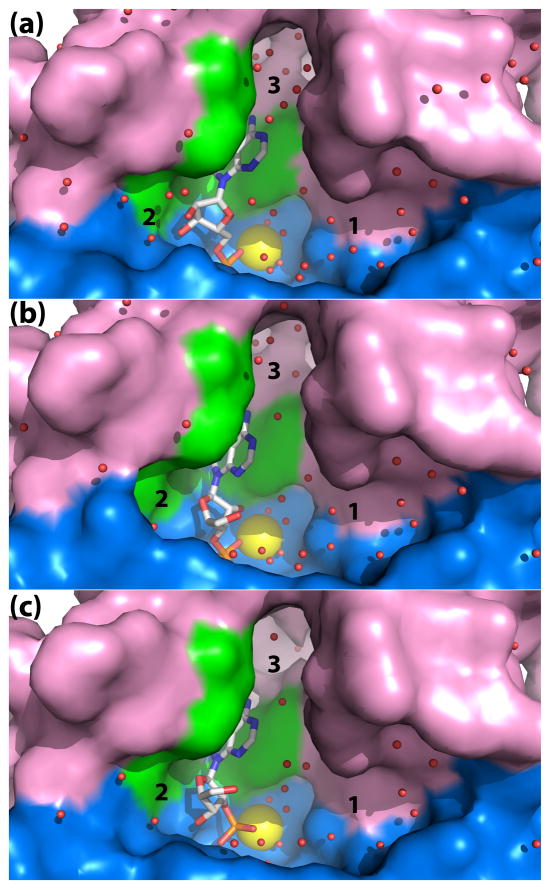
Fig. 3.
Close-up view of the rP4 active site emphasizing the shape and solvent content. The panels correspond to D66N complexed with (a) 5′-AMP, (b) 3′-AMP, and (c) 2′-AMP. Three solvent-filled pockets are labeled 1, 2, and 3. The core and cap domains are colored blue and pink, respectively, and residues of the aromatic box are colored green. The yellow sphere represents Mg2+.

