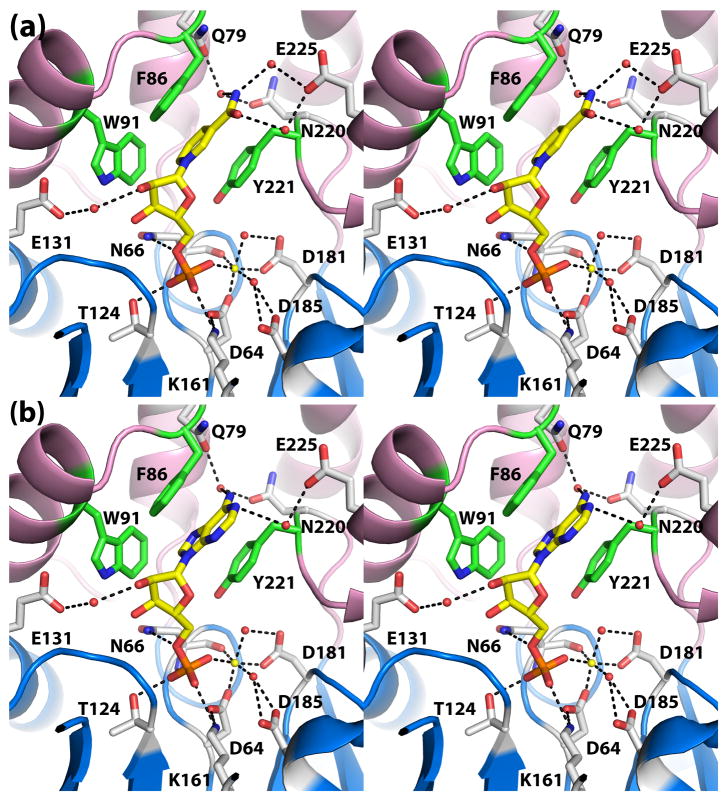
Fig. 4.
Recognition of the nucleoside 5′-monophosphate substrates (a) NMN and (b) 5′-AMP (stereographic views). In both panels, the substrate is represented in yellow sticks, and Mg2+ is depicted as a yellow sphere. Secondary structural elements of the core and cap domains are colored blue and pink, respectively, and residues of the aromatic box are colored green.

