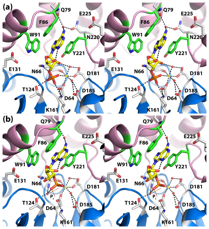
Fig. 5.
The active sites of D66N complexed with (a) 3′-AMP (b) 2′-AMP (stereographic views). In both panels, the substrate is represented in yellow sticks, and Mg2+ is depicted as a yellow sphere. Secondary structural elements of the core and cap domains are colored blue and pink, respectively, and residues of the aromatic box are colored green. In panel (b), the location of normal phosphoryl binding site is indicated by the phosphate ion shown in lines and labeled Pi.

