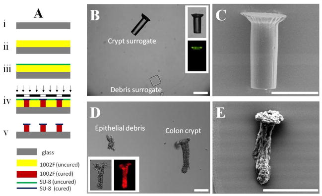
Fig. 1.
Surrogate crypts and colonic crypts (with debris). (A) Schematic of the fabrication process for the crypt surrogate. (B) Brightfield and fluorescence (inset) images of crypt surrogate. Debris surrogate is also shown. (C) SEM image of crypt surrogate. (D) DIC and brightfield/fluorescence (inset) images of isolated colon crypts. Typical epithelial debris is also shown. (E) SEM image of colon crypt. Scale bar is 50 μm.

