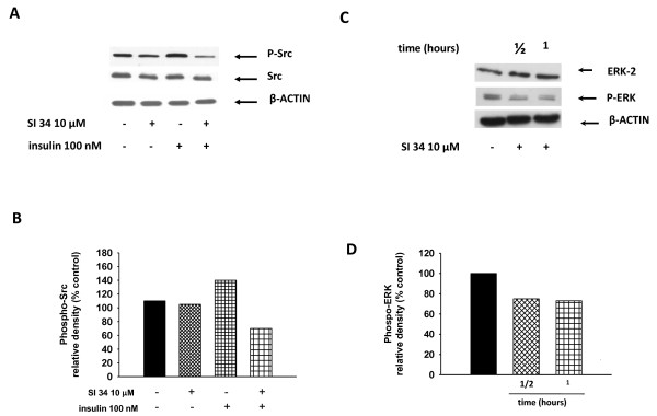
Figure 6.
Src- and ERK-phosphorylation in SH-SY5Y treated with SI 34. (A) SH-SY5Y cells were stimulated with insulin (100 nM; 30 min) in the presence or absence of SI 34 (10 μM; 30 min), and then western blotting analysis of Src and phospho-Src was performed. A Western blot, representative of three independent experiments showing similar results, is presented. (C) A representative gel (out of three) showed the ERK and phospho-ERK expression in presence or not of SI 34 is illustrated. (B and D) Densitometric analysis of immunoreactive bands corresponding to the Src-phosphorylated and ERK phosphorylated forms from blots A and C are reported. Autoradiographic bands were quantified by ImageJ software and normalized for β-actin levels. Data are reported as percentages of the values detected in untreated cultures.

