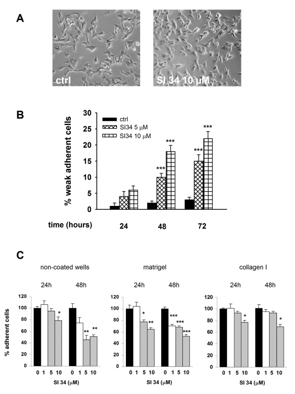
Figure 7.
SI 34 decreases SH-SY5Y cell adhesion. (A) Changes of cellular morphology in SH-SY5Y cultures exposed to 10 μM SI 34 for 72 hours. (B) Detached cells from cultures exposed (white bars) or not (black bars) to SI 34 were collected and counted as described in materials and methods. The results are expressed as percentage of detached cells (subtracted the percentage of dead cells from the full amount of detached cells) with respect to the total number of cells present in the well. Each value is the mean ± S.E.M. of 6 different sets of experiments made in triplicate. ***P < 0.001 vs respective controls. (C) Adhesion assay performed by plating SH-SY5Y cells on two different physiological substrates (Matrigel and collagen I) and on non-coated plastic surface for 30 min. Cells were treated with increasing concentrations of SI 34 (0, 1, 5, 10 μM) for 24 or 48 hours prior the adhesion assay. The values are expressed as mean percentage with respect to control (black bar) of at least three different measurements (± S.E.M). *, ** and ***P < 0.05, P < 0.01 and P < 0.001 vs respective control.

