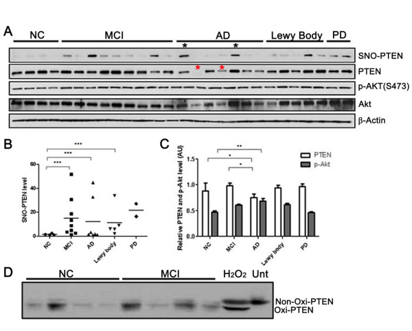
Figure 1.

Quantitative analysis of PTEN and P-Akt levels in relation to SNO-PTEN in human brains. (A) SNO-PTEN levels were detected by immunoblot analysis following biotin-switch assays. Other proteins were detected by Western blot analysis using 15 μg of brain lysate. (B) Quantification of the Western blots using densitometry analysis reveals a statistically significant elevation of SNO-PTEN levels in MCI and AD brains compared to NC brains *** indicates P < 0.001. (C) An inverse correlation between PTEN and p-Akt levels. * indicates P < 0.05 in MCI and AD compared to NC brains for total PTEN levels. ** indicates P < 0.005 between AD and NC groups for P-Akt. (D) Examination of H2O2-type oxidation of PTEN by band-shift assays: neuronal cells or brains were lysed in buffer containing 2% SDS and 40 mM N-ethylmaleimide and 20 μg of proteins was subjected to 10% SDS-PAGE under non-reducing condition as described [22]. "Unt" stands for untreated neurons. H2O2 was used at 100 μM for 2 h as a positive control for band-shift of the oxidized PTEN.
