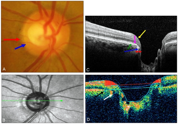
Fig. 3.
(A) Showed the extent of the β-zone (red arrow) and optic disc margin (blue arrow) on the temporal side of a healthy optic disc. (B) Peripapillary atrophy (PPA) was well-visualized by Spectralis optical coherence tomography (OCT) scanning laser ophthalmoscope imaging. (C) In a cross-sectional image of the optic disc scanned by the Spectralis OCT, the retinal nerve fiber layer (yellow arrow) and Bruch's membrane/retinal pigment epithelium complex layer (BRL) (green arrow) were observed in the β-zone PPA area, whereas the inner and outer segment complexes (pink arrow) were absent. The BRL edge showed slight posterior bowing around the optic disc margin. (D) The Stratus OCT image also showed slight posterior bowing of the BRL (white arrow), and the automatic disc margin detection algorithm failed to detect the edge of the optic disc margin (black arrow).

