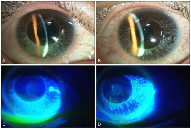
Fig. 1.
Slit-lamp photographs at the first examination (A,C, right eye; B,D, left eye). (A,B) Conjunctival injection, mild chemosis and deposition of the color pigments of the cosmetic contact lens onto the corneal epithelium were observed. The pattern of the deposition matched the pattern on the contact lenses. (C,D) Fluorescein-dye staining revealed punctate epithelial erosions and corneal epithelial defects in both eyes.

