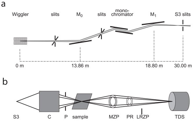
Fig. 1.
Typical transmission X-ray microscope setup (a) includes monochromator, and mirrors to focus beam on slits (S3). Within microscope (b) is a condenser (C) to focus beam onto sample, pinhole (P) to remove unfocused beam, x-y-z-θ sample stage with or without cryostat, micro zone plate (MZP), and optional phase ring (PR) for Zernike phase contrast. A low resolution zone plate (LRZP) is used to align the phase ring, and is removed for imaging. Transmission detector system (TDS) consists of scintillator to convert X-rays to visible light, objective and CCD detector. Diagrams are not to scale. Figure reproduced from Andrews et al. Micros. Microanal. (2010) 16: 327-336.

