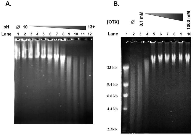
Figure 1. Establishment of OTX-coupled AGE assay.

(A) Examining pH effect on DNA alkaline denaturation. Genomic DT40 DNA was exposed to heat/acid conditions for 5 minutes to induce intact AP sites. The DNA was then exposed to various concentrations of NaOH to create a pH gradient (pH 10-13+) prior to being loaded into an agarose gel and electrophoresed under alkaline conditions (pH 12.4). The gels were stained with acridine orange and the image was obtained with a Kodak Image Station 440CF system. Lane 1 – No NaOH (Ø); Lanes 2-4 – pH ≤ 11.9; Lanes 5-8 – pH 12.2-12.6; Lanes 9-12 – pH ≥ 12.7. (B) OTX optimization for the protection of AP sites. Genomic DT40 DNA was exposed to heat/acid conditions for 5 minutes to induce intact AP sites. The DNA was then exposed to a concentration gradient of OTX (0.1-1000 mM) prior to denaturation with 100 mM NaOH (pH 12.8) and electrophoresed under alkaline conditions (pH 12.4) in an agarose gel. The gel was stained with acridine orange and the image was obtained with a Kodak Image Station 440CF system. Lane 1 – Marker; Lane 2 – No OTX (Ø); Lanes 3-10 – Heat/acid DNA + OTX (0.1, 0.3, 1, 3, 10, 30, 100 and 1000 mM).
