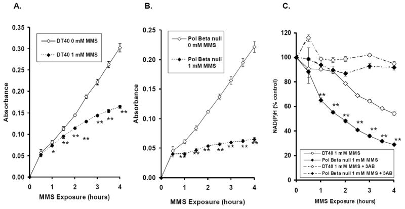
Figure 2. Intracellular NAD(P)H depletion in DT40 and Pol β-null cells exposed to 1 mM MMS or 1x PBS.

Intracellular NAD(P)H levels were detected by formation of formazan dye converted from tetrazolium salt in cells continuously exposed to 0 or 1 mM MMS for up to 4 hours. The amount of formazan dye in the culture medium was detected by absorbance at 450 nm using a plate reader. The intracellular NAD(P)H depletion is an indicator of PARP activation caused by accumulation of SSBs in the cell lines exposed MMS. (A-B) Absorbance measurements in the DT40 and Pol β-null cells, respectively. (C) DT40 and Pol β-null cells were continuously exposed to 1 mM MMS in the presence or absence of 3-AB (20 mM). Absorbance data plotted as percent control. The mean values represent three independent measurements. Bars indicate SD. Statistical Significance: * p < 0.05, ** p < 0.01, compared to PBS control (A-B) and DT40 cells (C) for the experiments in the absence of 3-AB.
