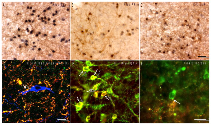
Fig. 1.

A–C: Colocalization of gonadal steroid receptors in kisspeptin neurons. Dual immunostained sections of sheep ARC showing high degree of colocalization of nuclear ER-α, PR and AR (blue-black) in kisspeptin cells (brown). Bar = 50 μm. D: Kisspeptin synaptic contacts onto a GnRH neuron. Confocal optical section (1 μm thick) of a triple-labeled section showing a terminal labelled with both kisspeptin (red) and synaptophysin (green) in direct contact with an ovine GnRH (blue) cell body (modified from Smith et al., 2008). Bar = 10 μm. E–F: Phenotypic heterogeneity between ARC and preoptic kisspeptin neurons. E: Colocalization of the endogenous opoid peptide, dynorphin (red), in kisspeptin neurons (arrows; green) of the sheep ARC. F: By contrast, kisspeptin neurons (green) in the sheep POA do not colocalize dynorphin even though they receive input from dynorphin-positive fibers (arrow; red). Bar = 20 μm. (modified from Goodman et al., 2007)
