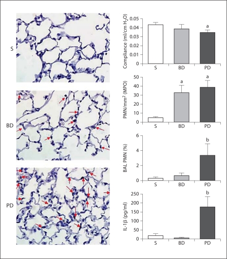
Fig. 3.
Representative photomicrographs (MPO staining) of mouse lung following 48 h of sham operation (S), BD ligation or PD ligation. Sham lungs were essentially normal morphologically. BD ligation or PD ligation resulted in vascular congestion with increased neutrophils (arrows). Graphs are described from top to bottom. Data are means ± SEM; a p < 0.05 vs. sham; b p < 0.05 vs. sham and BD ligation; ANOVA. Pulmonary compliance: after 24 h, the PD ligation group had decreased pulmonary compliance compared to sham, while with BD ligation compliance was not significantly reduced (n = 4–5/group). Morphometry of pulmonary PMN: after 48 h, the PMN count of MPO-stained lung sections in the PD ligation group was significantly higher than sham control, but was similar to the BD ligation group (MPO stain, n = 5 mice/group). BAL fluid PMN count: after 24 h, the BAL fluid showed an increased percentage of PMN after PD ligation compared to both controls (n ≥ 3/group). BAL fluid IL-1β concentration: after 48 h, ELISA of BAL fluid showed increased IL-1β concentration in the PD ligation group compared to both controls (n ≥ 3/group).

