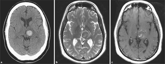
Fig. 2.
Brain imaging studies in a 60-year-old hypertensive male with a spontaneous left thalamic ICH. The CT diagnosis of hypertensive ICH (a) was changed after MRI review to ICH caused by a cavernous angioma (b, c). MRI shows a well-circumscribed lesion with a heterogenous reticulated core on the T2-weighted sequence (b) and an associated developmental venous anomaly with a ‘caput medusae’ appearance on the T1-weighted postcontrast sequence (c). This diagnosis was confirmed surgically.

