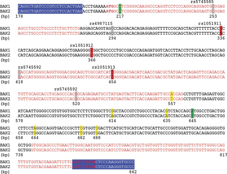
Figure 1.

Comparison of BAK1 cDNA (NCBI reference: NM_001188.3) and BAK2 genomic sequences (NCBI reference: NG_000850). The primers used by Gottlieb et al. [2009] for the RT-PCR of BAK1 are highlighted in blue. Sequences are numbered in base pairs using the same nomenclature as Gottlieb et al. [2009]. The three nucleotides highlighted in red are the three SNPs found variant by the authors in their original article, whereas the four remaining SNPs that they cited are highlighted in gray. The nucleotides highlighted in green are the two differences between BAK1 and BAK2 cited by Gottlieb et al. [2010] in their reply to Hatchwell [2010] in order to justify the absence of amplification of BAK2. The other differences between BAK1 and BAK2, which were not mentioned by Gottlieb et al., are highlighted in yellow.
